Neurologic Disorders - Assessment and Treatment of Balance
- Ana Souto
- Sep 25, 2023
- 6 min read
Updated: Jun 13, 2025
Part II - Balance Assessment with PhysioSensing
PhysioSensing has 12 assessment protocols that measure balance through COP displacement. Below, you can see our evidence-based recommendation of which PhysioSensing assessment protocols are adequate for each balance dimension within the neurologic disorder’s context.
Balance dimension | PhysioSensing Assessment |
Sensory reception and organization | mCTSIB, Romberg Test, Total Balance Pro |
Proprioception | Limits of Stability, Romberg Test, mCTSIB, Total Balance Pro |
Motor control | Limits of Stability, Rhythmic Weight Shift, Total Balance Pro, Sit to Stand |
Muscle strength | Weight Bearing Squat, Sit to Stand, Limits of Stability, Toral Balance Pro |
Reflexes and reaction time | Limits of Stability, Rhythmic Weight Shift, Sit to Stand, Total Balance Pro |
Postural control | Body Sway |
Weight distribution | Sit to Stand, Weight Bearing Squat |
Functionality | Sit to Stand |
How to improve with PhysioSensing?
After selecting the adequate assessment protocol for your patient, you can collect a substantial amount of objective data that will inform you about your patient balance status.
Let us create a scenario where you want to assess motor control, proprioception and have a functional assessment to complement. We are going to choose the following protocols: Romberg test, Limits of Stability and Sit to Stand.
Results
1. Limits of Stability Protocol

Clinical Report


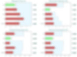
Limits of Stability Protocol can give us a lot of information about motor control, muscle strength and proprioception. What can we see in the results from our neurologic patient?
Reaction time: The results show a high reaction time, being the composite value (average of 8 directions) higher than the normative value. This can mean an apprehension/fear of losing balance, needing more time to prepare for the movement, or a dysfunction at cortical level causing a prolonged reaction time (Paraskevopoulou et al., 2021). Additionally, muscle weaknesses and rigidity could also partially explain it. These alterations are common in Parkinson’s Disease (Opara et al., 2017).
Movement Velocity: The results show a reduced movement velocity, being below the normative values. We can think about different causes for this. One that might be relevant is a reduction in proprioception, meaning that the patient needs more time to sense alterations in body position and to adjust to it in matters of muscle activation (neuromuscular activity), this may be caused by alterations in the mechanoreceptors at the soles of the feet, slowed transmission of somatic sensory impulses or changes in integration of sensory input (Melzer et al., 2008). Some of these alterations are common in polyneuropathy.
Endpoint Excursion and Maximum Excursion: The results show values below the normative data for the two parameters, except for movement in back direction. Two possible reasons for these might be lack of muscle strength on the plantar flexors, they need to support the leaning of the body to the front, and lack of ankle mobility or rigidity (Melzer et al., 2008). Another interesting finding in these results is the asymmetry between right and left direction. We will analyze this finding in association with the results from the Sit to Stand protocol.
Direction Control: The results show a decreased direction control, demonstrated by the composite (average) value. This means that an insufficient portion of the COP displacement happened in the wanted direction. The lack of direction control might signify problems in motor control, this can be due to alterations in afferent inputs, like proprioception, alterations in integration and organization of these inputs, and in the right muscle activation (neuro-muscular control). Another explanation for lack of direction control could be the presence of alterations in extra-pyramidal paths, we would need to clarify the cause, if it really is a sensory dysfunction or a cerebellum dysfunction. To do that we need to do the Romberg Test, if the increased body sway does not differ much between eyes open condition and eyes closed condition it might be related to cerebellum dysfunction (D’Angelo, 2018; Inojosa et al., 2020). In common with Endpoint Excursion and Maximum Excursion parameters, we can see an asymmetry between right-left movement and back-front movement in matters of direction control.
2. Sit to Stand Protocol

Clinical Report


While analyzing the results from the Sit to Stand Protocol we can see an asymmetry in COP displacement in the statokinesiograms, there is a tendency for mediolateral sway towards the left side, which is in accordance with the asymmetry seen during LOS assessment, additionally, the results also show a marked asymmetry between left and right weight distribution, this patient is clearly supporting more weight on the left leg. These are common findings in stroke and Parkinson´s patients, explained by lack of muscle strength and/or by a somatosensory deficit (De Nunzio et al., 2014; Frykberg & Häger, 2015). Moreover, an increase in weight time transfer has also been demonstrated in studies comparing stroke patients with healthy control subjects. (Chou et al., 2003).
3. Romberg test Protocol
Clinical Report


The Romberg test is commonly used for assessing balance, specifically the assessment of the dorsal column of the spinal cord that conducts proprioceptive sensory information. The test is positive if the patient loses balance with their eyes closed, this means increased body sway, placing one foot in the direction of the sway or falling. The foam surface will decrease the proprioceptive input, making the task more challenging. In the last condition, foam surface with eyes closed, the patient is relying mainly on the vestibular system input, so we can roughly assess the vestibular function (Inojosa et al., 2020). However, it is not a reliable indicator of vestibular function as we do need two systems input to maintain balance. There are specific tests for vestibular assessment on our Virtual Reality Software.
Looking into Romberg test results we can see an increase in COP displacement, in ellipse area, as well as in sway velocity during eyes closed condition. In addition, the Romberg Quotient also indicates a possible somatosensory deficit.The Romberg Quotient is calculated as the Ratio between closed and open eyes values.
4. Summary
These were the main findings from the 3 protocols used for the assessment of our neurological patient:
Probable proprioceptive deficit (Romberg test, LOS) |
Reduced limits of stability (LOS) |
Increased body sway in the medial lateral plane (Romberg test, Sit to Stand) |
Asymmetric weight transfer during voluntary movement in the medial lateral plane, in favor of the left side (Sit to Stand) |
Impaired directional control, possible deficit in motor control (LOS) |
Read part III to learn how to create a rehabilitation Protocol with PhysioSensing!
Do you have questions about this topic? Or want to know more about balance system?
Schedule a 15 minute talk with me!
Ana Souto

Meet Ana, a physiotherapist with a master's degree in human physiology, currently specializing in neurobiology. Her professional journey has led her to gain extensive expertise in both neurology and sports physiotherapy.
Ana currently serves as the clinical specialist at PhysioSensing, a cutting-edge Balance Assessment and training device. Leveraging her strong foundation in scientific research and evidence-based practices, Ana creates customized assessment and training plans. Her approach is firmly rooted in the latest scientific findings, ensuring that PhysioSensing users receive the most effective and up-to-date care.
In addition to her role in designing tailored programs, Ana plays a pivotal role in guiding new clients through the learning process of using PhysioSensing. She also provides advanced training and support to existing customers seeking to further deepen their clinical practice knowledge and stay on top of the latest scientific advancements.
6. References
Chou, S.-W., Wong, A. M. K., Leong, C.-P., Hong, W.-S., Tang, F.-T., & Lin, T.-H. (2003). Postural control during sit-to stand and gait in stroke patients. American Journal of Physical Medicine & Rehabilitation, 82(1), 42–47. https://doi.org/10.1097/00002060-200301000-00007
D’Angelo, E. (2018). Physiology of the cerebellum. In Handbook of Clinical Neurology (Vol. 154, pp. 85–108). Elsevier. https://doi.org/10.1016/B978-0-444-63956-1.00006-0
De Nunzio, A. M., Zucchella, C., Spicciato, F., Tortola, P., Vecchione, C., Pierelli, F., & Bartolo, M. (2014). Biofeedback rehabilitation of posture and weightbearing distribution in stroke: A center of foot pressure analysis. Functional Neurology, 29(2), 127–134.
Frykberg, G. E., & Häger, C. K. (2015). Movement analysis of sit-to-stand – research informing clinical practice. Physical Therapy Reviews, 20(3), 156–167. https://doi.org/10.1179/1743288X15Y.0000000005
Inojosa, H., Schriefer, D., Klöditz, A., Trentzsch, K., & Ziemssen, T. (2020). Balance Testing in Multiple Sclerosis-Improving Neurological Assessment With Static Posturography? Frontiers in Neurology, 11, 135. https://doi.org/10.3389/fneur.2020.00135
Melzer, I., Benjuya, N., Kaplanski, J., & Alexander, N. (2008). Association between ankle muscle strength and limit of stability in older adults. Age and Ageing, 38(1), 119–123. https://doi.org/10.1093/ageing/afn249
Opara, J., Małecki, A., Małecka, E., & Socha, T. (2017). Motor assessment in Parkinson`s disease. Annals of Agricultural and Environmental Medicine, 24(3), 411–415. https://doi.org/10.5604/12321966.1232774
Paraskevopoulou, S. E., Coon, W. G., Brunner, P., Miller, K. J., & Schalk, G. (2021). Within-subject reaction time variability: Role of cortical networks and underlying neurophysiological mechanisms. NeuroImage, 237, 118127. https://doi.org/10.1016/j.neuroimage.2021.118127